When you’re experiencing back pain, understanding its cause is often the first step toward finding relief. Dr. Jeffrey A. Moore uses various diagnostic tools, and among the most powerful is the Magnetic Resonance Imaging (MRI) scan. An MRI provides detailed images of your spine’s soft tissues, offering a comprehensive look that other imaging techniques might miss. However, like any valuable tool, an MRI has its strengths and limitations. This article aims to clarify what your MRI can and cannot tell you about your back pain.
Before diving into what an MRI reveals, let’s briefly understand the technology behind it.
How MRI Technology Functions
An MRI machine uses a powerful magnetic field and radio waves to create detailed images of organs and structures inside your body. Unlike X-rays or CT scans, an MRI does not use ionizing radiation. The magnetic field temporarily realigns hydrogen atoms, which are abundant in water molecules within your body. Radio waves are then pulsed, knocking these atoms out of alignment. When the radio waves are turned off, the hydrogen atoms return to their original alignment, releasing energy. This energy is detected by the MRI scanner and converted into detailed images by a computer.
Preparing for Your MRI
Typically, there’s little special preparation needed for an MRI. You may be asked to avoid food or drink for a few hours prior, especially if a contrast agent is used. It’s crucial to inform the technician about any metal implants in your body, such as pacemakers, artificial joints, or surgical clips, as these can interfere with the magnetic field or even pose a safety risk. You’ll likely be asked to remove all metal objects, including jewelry, before the scan. The scan itself involves lying still on a table that slides into a large tunnel-like machine. The machine can be noisy, and you may be offered earplugs.
In addition to understanding what your MRI can reveal about back pain, you may find it beneficial to explore the experiences of patients undergoing cervical disc replacement. This procedure is often discussed in relation to back pain management and recovery. For more insights on this topic, you can read the article on patient experiences and research priorities by visiting Cervical Disc Replacement: Patient Experience and More.
What an MRI Can Reveal About Your Back Pain
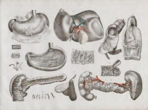
An MRI is excellent at visualizing soft tissues. This makes it invaluable for diagnosing a wide range of spine conditions that can contribute to back pain. Think of your spine like a complex building with many different materials. An X-ray might show the strong rebar (bones), but an MRI reveals the intricate wiring, plumbing, and insulation (discs, nerves, ligaments, muscles).
Disc Problems
The intervertebral discs are among the most common sources of back pain. An MRI provides a clear picture of these cushioning structures between your vertebrae.
- Herniated Discs: This occurs when the soft, gel-like center of a disc pushes through a tear in its tougher outer layer. An MRI can clearly show the extent of the herniation and if it’s pressing on nearby nerves. This pressure can lead to sciatica (pain radiating down the leg), numbness, or weakness.
- Bulging Discs: A bulging disc is when the disc extends beyond its normal boundaries but hasn’t completely ruptured. An MRI can identify the bulging and its proximity to nerve roots.
- Degenerative Disc Disease (DDD): As we age, discs can lose water content, become thinner, and develop small cracks. An MRI can show the typical signs of DDD, such as disc dehydration (appearing dark on the scan), loss of disc height, and osteophytes (bone spurs) that may develop as a consequence.
Nerve Compression
Many back pain symptoms stem from nerves being pinched or irritated.
- Spinal Stenosis: This is a narrowing of the spinal canal, which is the space that houses the spinal cord and nerve roots. Spinal stenosis can be caused by disc degeneration, bone spurs, thickened ligaments, or a combination. An MRI can precisely show the degree of narrowing and how it affects the neural structures.
- Radiculopathy: This term describes symptoms caused by a pinched nerve in the spine, often presenting as pain, numbness, tingling, or weakness radiating into an arm or leg. An MRI can pinpoint the exact location of the nerve compression.
Other Soft Tissue Issues
Beyond discs and nerves, an MRI can detect problems with other soft tissues and structures around the spine.
- Ligament Damage: Ligaments are strong bands of tissue that connect bones and stabilize the spine. An MRI can identify torn or strained ligaments that might be contributing to instability and pain.
- Muscle Injuries: While less common as a direct cause of chronic back pain, muscle strains or tears can be seen on an MRI, especially in cases of acute trauma.
- Spinal Cord Abnormalities: An MRI offers excellent visualization of the spinal cord itself, allowing for the detection of tumors, cysts, or other rare conditions affecting the cord.
- Inflammation or Infection: In some cases, back pain can be caused by inflammation (like in inflammatory arthritis) or infection within the spine (osteomyelitis, discitis). An MRI can show signs of inflammation and fluid collections, which are indicative of these conditions.
- Tumors: Although rare, tumors, both benign and malignant, can occur in the spine or surrounding tissues. An MRI is a crucial tool for detecting and characterizing these lesions.
- Scoliosis Evaluation: While X-rays are typically the primary tool for assessing the curvature of scoliosis, an MRI can be used to evaluate the underlying spinal cord for any abnormalities that might be contributing to or be associated with the spinal curve, especially in atypical or rapidly progressing cases.
- Trauma Follow-Up: After a spinal injury, an MRI can provide detailed information about the extent of damage to soft tissues, including ligaments, discs, and the spinal cord, complementing information gathered from X-rays or CT scans that focus on bone fractures. This helps guide treatment and prognosis.
What an MRI Can’t Tell You About Your Back Pain
While exceptionally powerful, an MRI is not a crystal ball. It has significant limitations, and these are crucial to understand to avoid misinterpretation and unnecessary anxiety. Just as a detailed architect’s blueprint tells you where the walls and pipes are, it doesn’t tell you the smell of fresh paint or the overall feeling of comfort a room might provide.
The Extent of Your Pain
Perhaps the most important limitation: an MRI cannot measure your pain. It shows structural abnormalities, but there’s often a disconnect between what the scan reveals and how much pain a person experiences.
- “Normal” Findings in Painful Spines: Many individuals with significant back pain have MRIs that show surprisingly few, or very minor, abnormalities. Their pain may be due to muscular issues, facet joint inflammation not clearly visible, or complex neurological factors.
- “Abnormal” Findings in Pain-Free Spines: Conversely, it’s very common for people with no back pain at all to have significant findings on an MRI, such as disc bulges, herniations, or degenerative changes. Imagine a person in their 40s or 50s. If you put 100 people without back pain into an MRI scanner, a large percentage would show some form of disc degeneration, bulging, or even herniation simply due to age and normal wear and tear. This highlights that structural findings don’t always equate to pain.
The Exact Cause of Your Pain (Always)
While an MRI can point to potential anatomical causes, it doesn’t always provide the definitive answer to why you are in pain.
- Multiple Potential Contributors: Many people have several MRI findings. Is the pain from the bulging disc, the facet joint arthritis, or a combination? The MRI shows you the possibilities, but clinical examination and patient history are vital for narrowing down the primary cause.
- “Non-Specific” Back Pain: A large percentage of back pain is classified as “non-specific,” meaning no clear structural cause can be identified even with advanced imaging. This doesn’t mean the pain isn’t real; it simply means the MRI isn’t showing the underlying issue, which could be related to muscle imbalance, poor posture, stress, or other factors not visible on the scan.
The Need for Surgery
An MRI alone never dictates the need for surgery. It provides an anatomical road map, but your symptoms, response to conservative treatments, and a thorough clinical evaluation are far more important in deciding on surgical intervention.
- Conservative Care First: Many significant MRI findings, even herniated discs, can resolve or become asymptomatic with time and conservative care (physical therapy, medications, injections). Dr. Moore emphasizes exploring these less invasive options first.
- Surgery for Symptoms, Not Scans: Surgery is typically considered only when severe or persistent symptoms correlate directly with an MRI finding and have not improved with conservative management. For example, a large disc herniation that is causing progressive weakness in your leg, despite several months of physical therapy, would be a strong candidate for surgery.
The Biomechanical Impact
An MRI provides static images. It shows the spine in a fixed position. It can’t fully capture how dynamic movements, posture, or specific activities impact your pain.
- Movement-Related Pain: Many people experience pain that is exacerbated by certain movements or positions but not others. While an MRI shows the structures, it doesn’t explain the biomechanical stresses at play during these movements.
- Muscle Function: An MRI can show muscle tears but doesn’t assess muscle strength, endurance, or how muscles are firing (or not firing) during movement, which are often key contributors to chronic back pain.
Diagnosing Your Back Pain: A Holistic Approach
When you consult Dr. Moore for back pain, the MRI is just one piece of the puzzle. He employs a comprehensive diagnostic approach.
The Clinical Examination
This is perhaps the most important part of the diagnostic process. Dr. Moore will ask you detailed questions about:
- Your symptoms: When did the pain start? Where is it located? What does it feel like (sharp, dull, burning)? Does it radiate?
- Aggravating and alleviating factors: What makes the pain worse or better?
- Medical history: Any previous injuries, surgeries, or underlying conditions.
- Lifestyle: Your occupation, activity level, and habits.
He will then perform a physical examination, assessing your range of motion, muscle strength, reflexes, and sensation. He’ll look for specific signs that point to nerve irritation or other issues.
The Role of X-rays
While an MRI excels at soft tissues, X-rays are superior for visualizing bones. They can show:
- Bone alignment: Such as with scoliosis or spondylolisthesis (one vertebra slipping over another).
- Fractures: Breaks in the bones.
- Bone spurs (osteophytes): Which can narrow the spinal canal.
- Overall spinal stability: Sometimes taken in different positions (flexion/extension) to see how the spine moves.
Often, both X-rays and an MRI are used together to provide a complete picture of your spinal health.
In exploring the complexities of back pain diagnosis, it is essential to consider various advancements in medical technology that can aid in treatment. A related article discusses the latest innovations in spine orthopedic medtech, highlighting how companies like Medtronic and Orthofix are revolutionizing patient care. You can read more about these developments and their implications for back pain management in this insightful piece. For further information, check out the article here.
Safety Red Flags: When to Seek Immediate Attention
| Aspect | What MRI Can Tell You | What MRI Can’t Tell You |
|---|---|---|
| Structural Abnormalities | Detects herniated discs, spinal stenosis, tumors, infections, and fractures | Cannot confirm if these abnormalities are the source of pain |
| Disc Degeneration | Shows disc dehydration, bulging, or degeneration | Does not correlate well with pain severity or presence |
| Nerve Compression | Identifies nerve root impingement or inflammation | Cannot always predict if nerve compression causes symptoms |
| Soft Tissue Evaluation | Visualizes muscles, ligaments, and other soft tissues | May not detect subtle muscle strain or functional issues |
| Functional Information | Provides detailed anatomical images | Does not provide information on pain mechanisms or functional impairment |
| Incidental Findings | May reveal abnormalities unrelated to pain | Can lead to unnecessary worry or interventions |
While most back pain is not an emergency, there are certain “red flag” symptoms that warrant immediate medical evaluation. If you experience any of these, contact Dr. Moore’s office or seek emergency care promptly:
- Sudden onset of severe weakness in your legs or arms.
- Difficulty controlling your bladder or bowels (urinary or fecal incontinence, or retention).
- Numbness in the “saddle area” (buttocks, groin, inner thighs), also known as saddle anesthesia.
- Unexplained weight loss.
- Fever or chills accompanying back pain.
- Back pain that is constant, progressive, and not relieved by rest, especially at night.
- Back pain following a significant trauma (e.g., fall, car accident).
- New or worsening neurological symptoms (e.g., foot drop, severe radiating pain).
If you’re looking to understand more about the complexities of back pain and its treatment options, you might find it helpful to read about the potential benefits of semaglutide in managing complications for lumbar spine patients. This related article provides insights into how this medication could play a role in improving outcomes for those suffering from back issues. You can check it out here.
Treatment Paths: From Conservative Care to Advanced Surgery
Once a diagnosis is made, Dr. Moore specializes in tailoring a treatment plan specific to your needs, always prioritizing the least invasive yet most effective options.
Conservative Treatment Options
Most cases of back pain improve with conservative management. Here are some common approaches:
- Physical Therapy: Targeted exercises to strengthen core muscles, improve flexibility, correct posture, and reduce pressure on nerves.
- Medications: Over-the-counter pain relievers (NSAIDs), muscle relaxants, or neuropathic pain medications prescribed by your physician.
- Injections: Epidural steroid injections can reduce inflammation around spinal nerves, providing temporary pain relief. Facet joint injections or nerve blocks can also be used diagnostically and therapeutically.
- Lifestyle Modifications: Weight management, ergonomic adjustments at work, regular low-impact exercise, and smoking cessation.
- Chiropractic Care/Osteopathic Manipulation: For some, these treatments can help with alignment and mobility.
Surgical Interventions
When conservative treatments fail to provide adequate relief, and symptoms are persistent or worsening, Dr. Moore offers advanced surgical solutions. He specializes in minimally invasive approaches whenever appropriate, which can lead to faster recovery times and less post-operative pain.
- Minimally Invasive Decompressions:
- Microdiscectomy: A small portion of a herniated disc is removed to relieve pressure on a nerve.
- Laminectomy/Laminotomy: A small part of the vertebral bone (lamina) is removed to enlarge the spinal canal and relieve pressure on nerves, often employing ultrasonic decompressions for precision and reduced tissue damage.
- Foraminotomy: Enlarging the opening (foramen) where a nerve root exits the spinal canal.
- ProneTransPsoas (PTP): A revolutionary minimally invasive approach for lumbar fusion that accesses the spine through the side of the body, allowing for precise disc removal and cage placement while minimizing disruption to posterior muscles and structures. This can be key for addressing certain disc issues and stabilizing the spine.
- Fusion Procedures:
- Spinal Fusion: Two or more vertebrae are permanently joined together to stabilize a painful or unstable segment of the spine. This can be performed in the cervical (neck), thoracic (upper back), or lumbar (lower back) regions using various approaches (anterior, posterior, lateral, or transforaminal).
- Interbody Fusions (e.g., ALIF, PLIF, TLIF, XLIF, PTP): These involve removing the disc and placing a bone graft and/or a cage between the vertebrae to promote fusion, often accompanied by screws and rods for immediate stability. Dr. Moore is proficient in these techniques, including the advanced PTP approach.
- Disc Replacement:
- Arthroplasty: For selected patients, artificial disc replacement may be an option, particularly in the cervical spine. This procedure removes the damaged disc and replaces it with an artificial one, designed to preserve motion at that spinal segment, unlike fusion.
- Scoliosis Correction:
- For adults with significant pain or progressive curvature due to scoliosis, surgical correction may involve a combination of fusion, instrumentation (rods and screws), and sometimes osteotomies (bone cuts) to realign the spine and alleviate symptoms.
- Trauma Follow-up:
- After initial management of spinal fractures or dislocations, ongoing care may involve surgery to stabilize the spine, decompress neural structures, or manage complications arising from the trauma.
Frequently Asked Questions
Q: Is an MRI always necessary for back pain?
A: No. In many cases, back pain improves with conservative care without the need for an MRI. Imaging is generally reserved for cases where pain persists, worsens, or is accompanied by “red flag” symptoms.
Q: Why do I need to lie still during an MRI?
A: Any movement during the scan can blur the images, making them difficult to interpret accurately.
Q: Can an MRI show soft tissue damage that an X-ray misses?
A: Yes, absolutely. X-rays are best for bones, while MRIs are superior for visualizing soft tissues like discs, nerves, ligaments, and the spinal cord.
Q: Will my insurance cover an MRI?
A: Most insurance plans cover medically necessary MRIs. It’s always best to check with your insurance provider directly to understand your specific benefits and any pre-authorization requirements.
Q: How long does an MRI take?
A: A typical spine MRI can take anywhere from 20 to 60 minutes, depending on the specific areas being scanned and the number of sequences required.
Take the Next Step for Your Back Health
Understanding your MRI is just one part of your journey to finding relief from back pain. Dr. Jeffrey A. Moore believes in empowering you with knowledge and providing compassionate, expert care. If you are experiencing persistent back or neck pain, or if you have an MRI you would like a second opinion on, don’t hesitate to reach out.
We invite you to call us at (405) 645-5475 to schedule an appointment or book online at JeffreyMooreSpine.com. We also offer a FREE MRI review/2nd opinion service to help you understand your options without obligation. Your journey to a healthier spine starts here.
FAQs
What is an MRI and how is it used to diagnose back pain?
An MRI (Magnetic Resonance Imaging) is a non-invasive imaging technique that uses magnetic fields and radio waves to create detailed images of the spine, discs, nerves, and surrounding soft tissues. It helps doctors identify structural abnormalities that may be causing back pain.
Can an MRI always determine the exact cause of back pain?
No, an MRI cannot always pinpoint the exact cause of back pain. Many people have abnormalities on MRI scans, such as disc bulges or degenerative changes, without experiencing any pain. Clinical correlation with symptoms and physical examination is essential.
What types of back problems can an MRI detect?
An MRI can detect herniated discs, spinal stenosis, nerve compression, tumors, infections, fractures, and degenerative disc disease. It provides detailed images of soft tissues, which X-rays cannot show.
Are there limitations to what an MRI can reveal about back pain?
Yes, MRIs cannot show functional issues like muscle strain, inflammation, or pain caused by non-structural factors. They also cannot measure pain intensity or differentiate between painful and non-painful abnormalities.
Is an MRI always necessary for someone with back pain?
No, MRIs are not always necessary, especially in cases of acute back pain without red flags (such as severe neurological symptoms or suspicion of serious conditions). Many cases improve with conservative treatment without imaging.
Can an MRI result lead to unnecessary treatments or surgeries?
Yes, incidental findings on MRI can sometimes lead to overdiagnosis and overtreatment, including unnecessary surgeries. It is important to interpret MRI results in the context of clinical symptoms.
How should MRI findings be interpreted in the context of back pain?
MRI findings should be combined with a thorough medical history, physical examination, and other diagnostic tests. A healthcare professional can determine whether the abnormalities seen on MRI are likely related to the patient’s symptoms.
Are there risks associated with getting an MRI for back pain?
MRI is generally safe and does not use ionizing radiation. However, it may not be suitable for patients with certain implants, pacemakers, or metal fragments in the body. Some patients may experience claustrophobia during the scan.
What other diagnostic tools are used alongside MRI for back pain?
Other tools include X-rays, CT scans, electromyography (EMG), nerve conduction studies, and physical examinations. These help provide a comprehensive assessment of back pain causes.
How can patients best prepare for an MRI scan?
Patients should inform their doctor about any metal implants, allergies, or pregnancy. They should remove all metal objects before the scan and follow any specific instructions provided by the imaging center.